Scientists Rebuilt a 3.67-Million-Year-Old Face, and It Didn’t Look the Way Anyone Imagined
For decades, scientists have studied ancient fossils to better understand how early human relatives evolved across the African continent. One of the most fascinating discoveries in this field is the skeleton known as “Little Foot,” a remarkably complete hominin fossil uncovered in South Africa. Recent technological advances have allowed researchers to digitally reconstruct this ancient individual’s face for the first time, offering intriguing clues about the connections between early human populations that lived millions of years ago.
Revealing The Story Behind Little Foot
More than 3.6 million years ago, a small-brained hominin walked upright through landscapes that today form part of South Africa. This individual, nicknamed “Little Foot,” represents one of the most complete early hominin skeletons ever discovered. Because of its exceptional preservation, the fossil has become a key piece of evidence for understanding how early members of the human lineage lived and evolved long before modern civilization emerged.
A Fossil That Sparked Scientific Debate

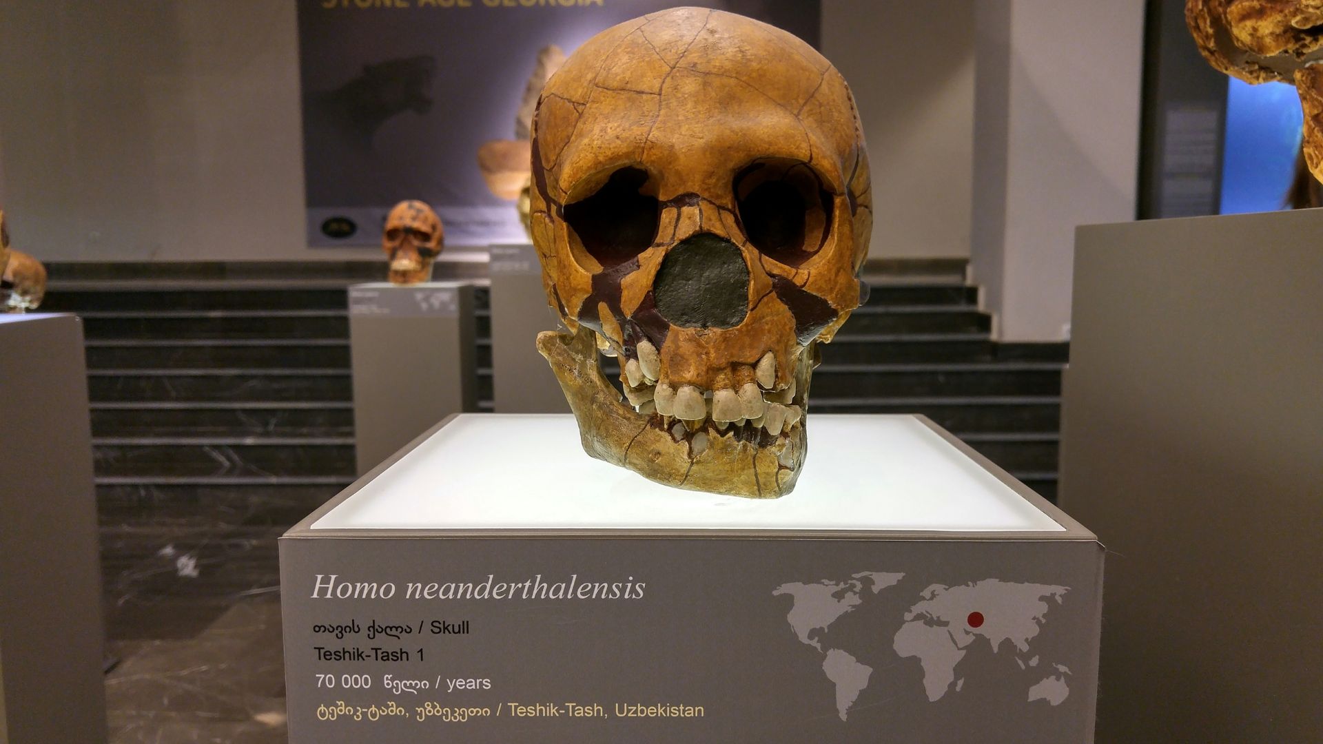
Little Foot has long captured the attention of paleoanthropologists because it dates to roughly 3.67 million years ago, making it the oldest known hominin from southern Africa. Many researchers classify it as Australopithecus prometheus, a member of the broader Australopithecus genus that existed before the emergence of Homo. Studying its anatomy may help scientists determine whether early human relatives across Africa were closely connected populations or groups that evolved separately over time.
Reconstructing A Face Lost For Millions Of Years
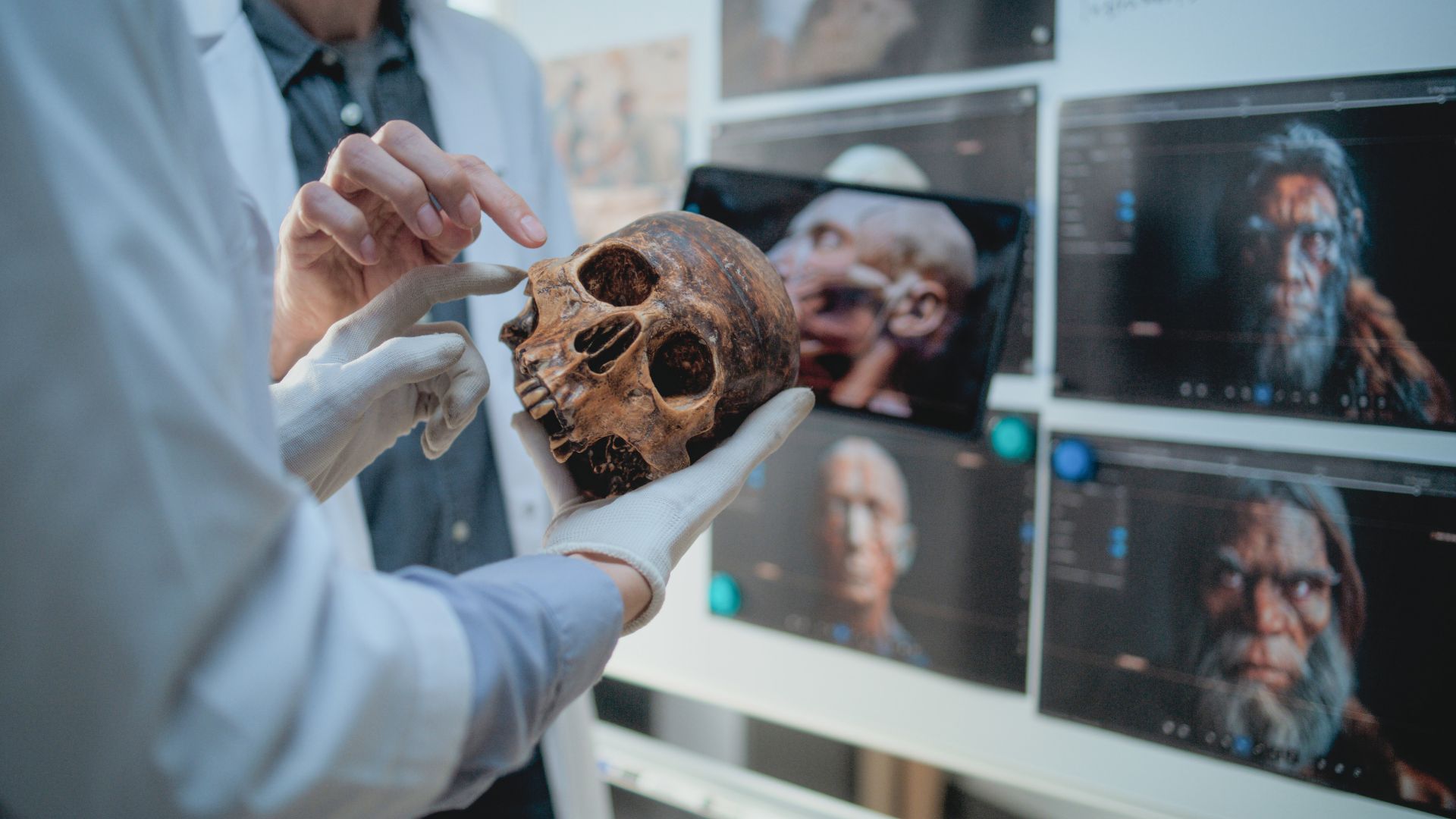
The skull of Little Foot endured immense geological pressure during its long burial inside Sterkfontein Cave, causing the facial bones to shift, crack, and distort. When researchers completed the painstaking excavation of the skeleton in 2016, the face was fragmented and difficult to interpret. Rebuilding it required advanced imaging technology and digital tools capable of carefully reconstructing the original structure of the fossilized bones.
The Role Of Powerful Particle Accelerators

To achieve this reconstruction, scientists brought the fossil to the Diamond Light Source in the United Kingdom, a synchrotron facility that produces highly intense X-rays. Unlike conventional medical scanners, this technology can penetrate dense rock and fossilized bone, capturing extremely detailed images. Using these beams, researchers produced thousands of cross-sectional scans that revealed hidden structures within the skull.
Turning Scans Into A Digital Reconstruction

Specialized software allowed researchers to separate bone fragments from surrounding rock in the digital scans. By analyzing the structure layer by layer, the team identified several major bone sections that had shifted from their original positions. These pieces were carefully repositioned in a virtual model, while the damaged left side of the face was reconstructed by mirroring the preserved right side.
Comparing Little Foot With Other Primates

Once a full digital face was reconstructed, researchers compared it with skulls from modern primates such as gorillas, chimpanzees, orangutans, and humans. They also examined two well-known Australopithecus fossils, Sts 5 from South Africa and A.L. 444-2 from Ethiopia. By mapping dozens of anatomical reference points across each skull, scientists measured several facial dimensions to understand how Little Foot fit within the evolutionary landscape.
Unexpected Similarities With Eastern African Fossils
The results revealed an intriguing pattern. In several digital shape analyses, Little Foot’s reconstructed face showed closer similarities to the Ethiopian fossil A.L. 444-2 than to Sts 5, another specimen discovered in the same South African region. This observation suggests that certain facial characteristics may have been shared across geographically distant Australopithecus populations.
Distinctive Features In Facial Structure

Little Foot’s face appears relatively large compared with other fossils from the same genus, with measurements that fall within ranges seen in large primates such as gorillas and orangutans. The shape of the eye sockets is also notable, appearing more oval, similar to those of orangutans and the Ethiopian fossil. In contrast, Sts 5 displays more rectangular orbital shapes, highlighting anatomical differences that could reflect evolutionary changes over time.
Implications For Early Human Evolution

These findings raise the possibility that early Australopithecus populations across eastern and southern Africa shared certain facial patterns during the Pliocene epoch, a geological period that began about five million years ago. If confirmed, this could indicate broader biological connections between these populations, though researchers emphasize that the evidence is still limited and open to interpretation.
A Reconstruction That Opens New Questions

Despite the excitement surrounding the reconstruction, scientists stress that the current model represents an early step in the research process. The digital assembly of bone fragments did not yet correct for distortions caused by fossilization over millions of years. Future studies using advanced retrodeformation techniques may refine the facial structure further, potentially revealing even deeper insights into the evolutionary story behind Little Foot.